


Literature Review | Methodological Validation and Clinical Value of Cellgenebio Flow Fluorescence Technology in AD Blood Detection
Blood biomarker detection for Alzheimer's disease (AD) is entering a new phase of clinical application. Since 2024, multiple domestic and international guidelines have incorporated plasma P-tau217 and P-tau181 as core indicators in AD diagnosis and management. However, mainstream detection platforms largely rely on specialized equipment with high costs, limiting their adoption in primary care and large-scale screening. Flow fluorescence technology (CBA), as a widely accessible open platform, offers a new approach to address this bottleneck.

Cellgenebio has developed the world's first AD blood biomarker multiplex detection kit (flow fluorescence luminescence method) based on this technology, which has accumulated clinical validation results from multiple research teams. In terms of diagnostic performance, when benchmarked against the clinical "gold standard" (Aβ-PET imaging), plasma P-tau181 achieved an AUC of 0.904; the P-tau181/Aβ42 ratio improved diagnostic performance to over 95%, with results highly consistent with the Simoa platform. In a head-to-head comparison of three novel P-tau217 detection technologies, flow fluorescence technology showed no significant difference in diagnostic performance compared to single-molecule immunoassay technology, with specificity reaching 100%. Furthermore, this technology has been successfully applied to exclude AD pathology in cerebral small vessel disease research, ensuring cohort purity. These methodological validations and clinical data demonstrate that the Cellgenebio flow fluorescence platform delivers reliable performance, providing an accessible and cost-effective detection pathway for AD early screening and differential diagnosis.

This article systematically analyzes the methodological characteristics and clinical application value of Cellgenebio's flow fluorescence technology in AD detection, based on four published studies.
Literature 1 [1]: Diagnostic Performance of Flow Fluorescence Technology for Measuring Plasma P-tau181, Aβ40, and Aβ42
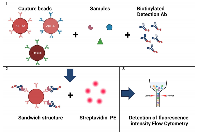
This study enrolled a total of 50 patients with cognitive impairment (CI) and 22 healthy controls (CU). Based on amyloid (Aβ) PET scan results, CI patients were divided into Aβ-positive and Aβ-negative groups. Plasma levels of Aβ40, Aβ42, and P-tau181 were measured in all participants using flow fluorescence technology, and cerebrospinal fluid (CSF) samples from 28 CI patients were also tested. All detection reagents were provided by Hangzhou Cellgene Biotech Co., Ltd.
Study results:
1. Plasma P-tau181: Accurately Distinguishes AD Patients
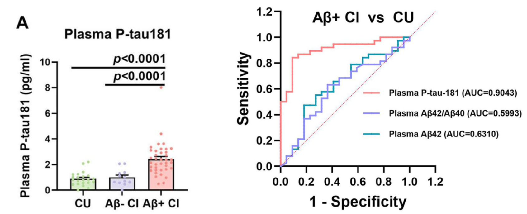
Plasma P-tau181 levels in Aβ PET-positive cognitive impairment (Aβ+CI) patients were significantly higher than those in cognitively normal individuals (CU) and Aβ PET-negative cognitive impairment (Aβ-CI) patients. Its diagnostic accuracy (AUC = 0.9043) far exceeded that of the conventional Aβ42/40 ratio. This indicates that detecting plasma P-tau181 via flow fluorescence technology can efficiently identify AD-related pathological features.
2. Plasma P-tau181: Dynamically Reflects Disease Progression
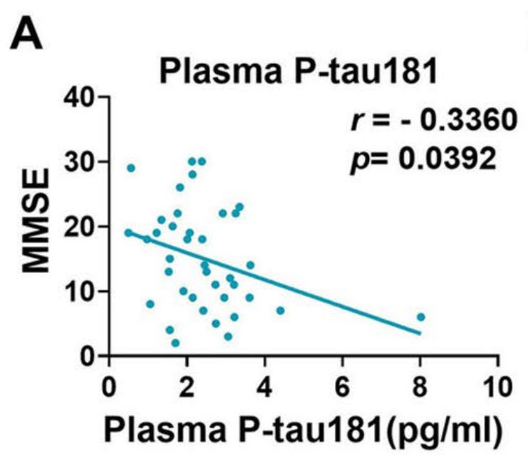
In addition to accurately identifying AD pathological changes, plasma P-tau181 levels showed a significant negative correlation with MMSE scores (r = -0.336), suggesting that it can dynamically reflect disease deterioration.
3. High Correlation Between Blood and CSF Results
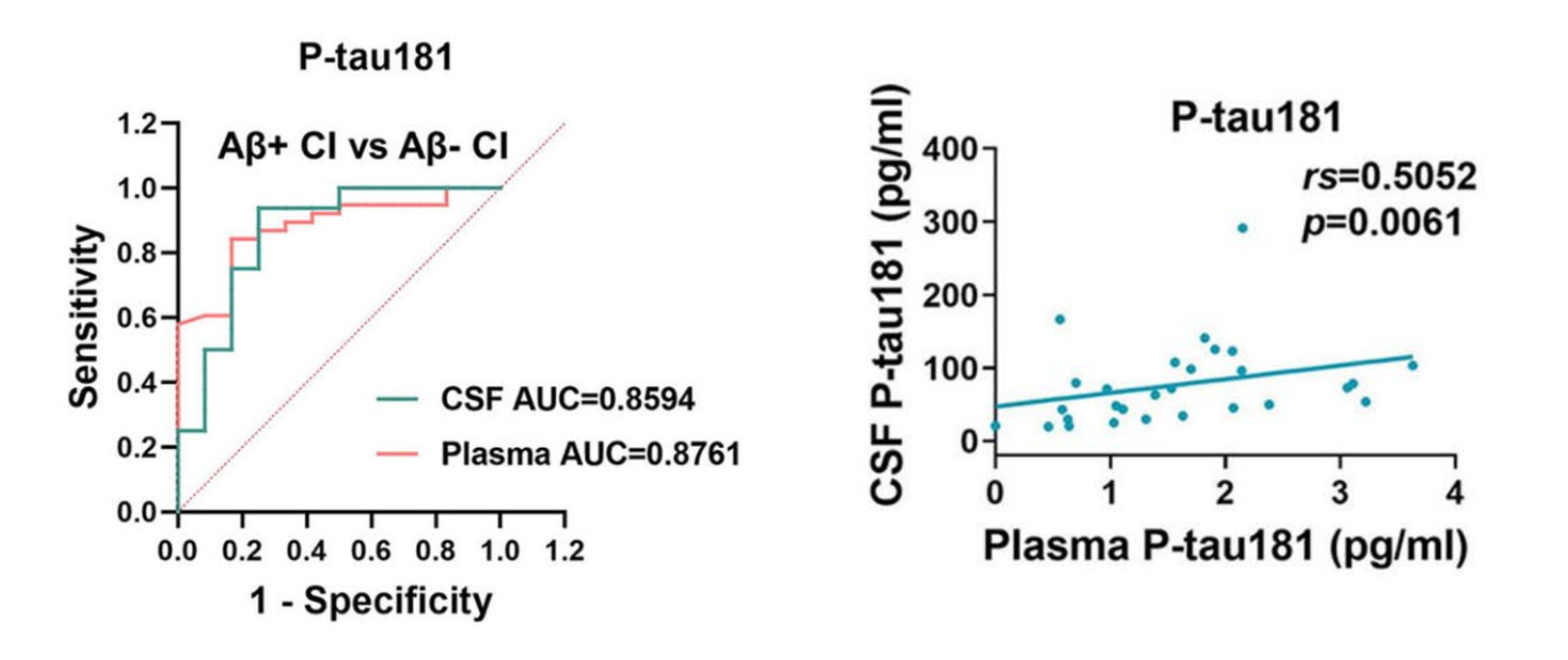
Among 28 CI patients, plasma and CSF P-tau181 levels showed a significant positive correlation (rs = 0.5052), and both demonstrated similar discriminative ability for Aβ+CI patients (AUC of 0.8761 and 0.8594, respectively), confirming the reliability of blood-based testing.
4. Flow Fluorescence Technology: Low Cost and High Feasibility
Based on flow fluorescence technology, multiple biomarkers can be detected simultaneously with only a minimal sample volume (25 μl). The platform is open and accessible, with costs far lower than Simoa, MSD, and other technologies, making it suitable for clinical adoption and of significant value in the early diagnosis, differential diagnosis, and monitoring of AD.
Literature 2 [2]: A High-Precision, Low-Cost AD Blood Test: Validating P-tau181/Aβ42 in a Real-World Cohort
This study conducted a comprehensive analysis of data from two cohorts (Cohort I included patients recruited from the memory clinic of Tianjin Medical University General Hospital; Cohort II was derived from a study on AD in the Chinese population conducted at Xuanwu Hospital). The final cohort comprised 123 controls, 60 AD/MCI patients, 34 FTD cases, and 34 SIVD patients, with AD patients meeting the International Working Group (IWG)-2 criteria and testing positive for Aβ PET.
Study results:
Plasma P-tau181/Aβ42 measured by flow fluorescence technology demonstrated robust diagnostic performance in distinguishing AD patients from healthy individuals in a clinical setting, with an accuracy of 96.2% (95% CI: 93.4%–99.0%), sensitivity of 95% (85.2%–98.7%), and specificity of 96.7% (91.4%–99.0%). It also showed a clear advantage in differentiating AD from non-AD dementias (SIVD, FTD) (see Table 1).
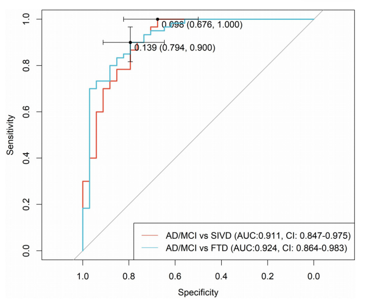
Figure: ROC Analysis for AD/MCI vs. SIVD and AD/MCI vs. FTD
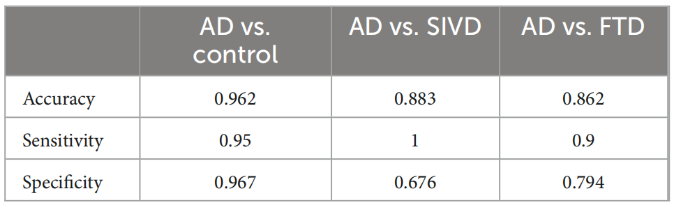
Table 1: Diagnostic Performance Evaluation of P-tau181/Aβ42 on the Flow Cytometry Platform
Furthermore, to assess the diagnostic concordance between P-tau181/Aβ42 and P-tau217, the study also performed Simoa P-tau217 testing on the samples. In a subset of participants (94 individuals; 38 AD/MCI, 56 controls), P-tau181/Aβ42 (flow cytometry) and Simoa P-tau217 showed an overall agreement of 88.3% (Table 2), and both methods demonstrated relatively similar diagnostic performance. This finding indicates that these two biomarkers are mutually corroborative in AD diagnosis, underscoring the strong reliability of plasma P-tau181/Aβ42 measured by flow cytometry as an AD diagnostic biomarker.
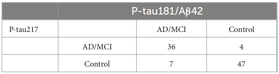
Table 2: Diagnostic Concordance Between P-tau181/Aβ42 (Flow Cytometry Platform) and P-tau217 (Simoa Platform)
Literature 3 [3]: Diagnostic Performance of Different Detection Methods for Plasma P-tau217 in AD
This study evaluated and compared the diagnostic performance of three novel plasma P-tau217 detection methods in a Chinese population cohort [DiSMS (single-molecule immunoassay), Ly Medivh™ AXL (single-molecule immunoassay), and CBA (Cellgenebio flow fluorescence method)], using ALZpath Simoa as the reference method. A total of 233 subjects were enrolled, all from the longitudinal study cohort at Tianjin Medical University General Hospital: 39 cognitively normal controls (CUC) and 194 cognitive impairment patients (including 28 MCI due to AD, 57 ADD, 70 SIVD, and 39 FTLD).
Key results:
1. Diagnostic Performance (ROC-AUC): Flow Fluorescence Technology on Par with Single-Molecule Immunoassay
DeLong test: Showed no statistically significant differences in AUC among the three methods (P > 0.05), indicating that flow fluorescence detection technology performs comparably to emerging single-molecule immunoassay technologies in diagnosing dementia-stage AD.
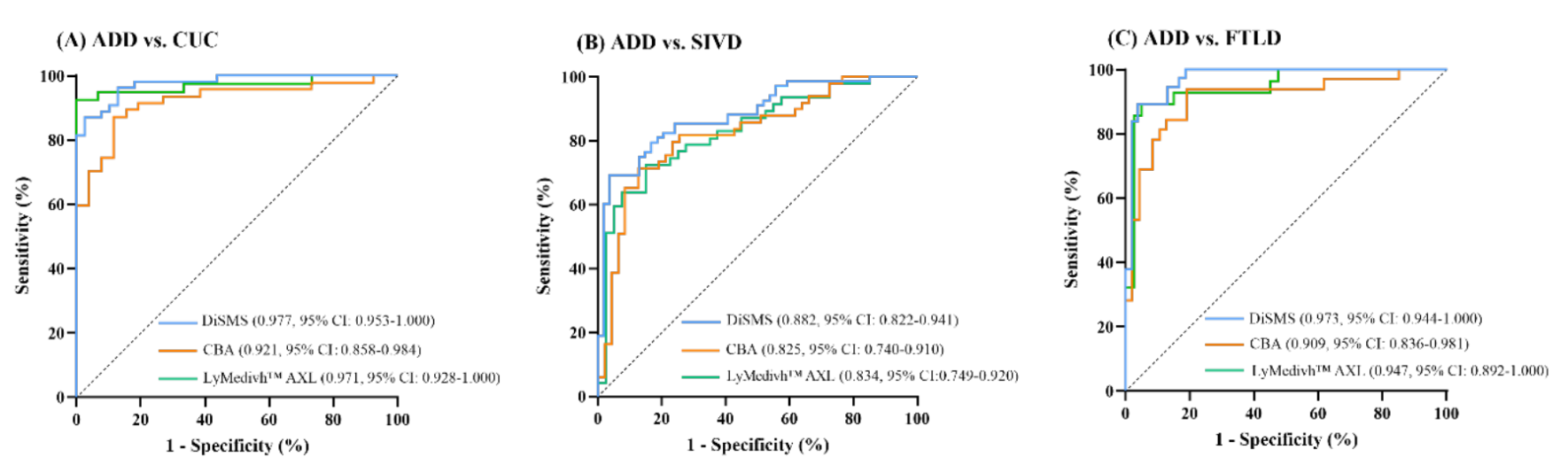
Figure: Performance of Four Detection Methods for Plasma P-tau217 in Distinguishing AD Dementia Patients from Controls (CUCs)
2. Diagnostic Metrics at Optimal Cut-off Values (ADD vs. CUC)
All four methods achieved 100% specificity and positive predictive value, suggesting high reliability of positive results and suitability for clinical trial enrollment screening and disease-modifying therapy (DMT) patient selection.
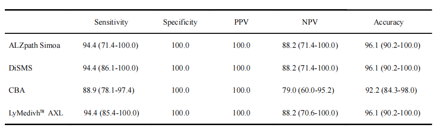
Table 1: Performance of Four Detection Methods for Plasma P-tau217 in Distinguishing AD from CUCs
3. Differences in Plasma P-tau217 Levels Across Groups
All three methods consistently showed:
P-tau217 levels in the ADD group were significantly higher than those in the CUC, SIVD, and FTLD groups (p < 0.05). Notably, the CBA method also revealed that ADD group levels were higher than the MCI group (p < 0.05), a difference not detected by the other two methods, suggesting that flow fluorescence detection has the capability to identify early pathological changes.
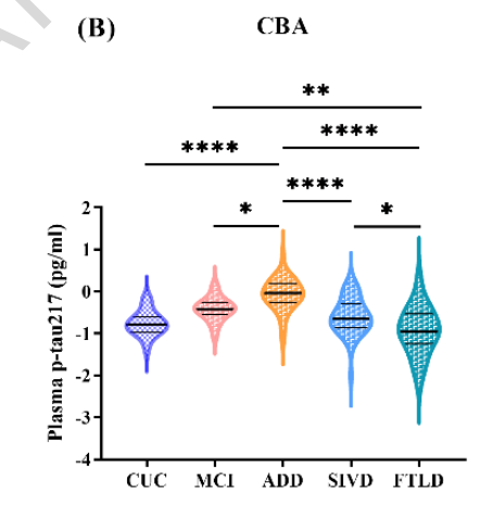
Figure: Validation of Between-Group Differences in Plasma P-tau217 Measured by Flow Fluorescence Method
Literature 4 [4]: Exclusion Diagnostic Value of CBA Technology in CSVD-Related MCI Research
This study systematically excluded cerebral small vessel disease (CSVD) patients with concomitant AD pathology through plasma biomarker detection, ensuring the "CSVD purity" of the study cohort. This design reflects the exclusion diagnostic value of flow fluorescence detection technology in non-AD dementia research: effectively distinguishing pure CSVD from CSVD with AD pathology, avoiding the confounding effect of AD pathology on cognitive impairment assessment in CSVD, and providing a pure CSVD sample for retinal-cerebral microvascular association studies.
Conclusion
Currently, mainstream AD blood detection platforms each have their own characteristics, and flow fluorescence technology, with its unique advantages, provides an alternative methodological choice for AD biomarker detection with high accessibility. This technology leverages flow cytometers already equipped in secondary and tertiary hospitals nationwide as an open platform, featuring high equipment accessibility, strong multiplex detection capability, low sample volume requirements, and excellent cost-effectiveness, making it particularly suitable for large-scale screening and routine clinical testing.
The aforementioned studies confirm that flow fluorescence technology demonstrates clear clinical value in scenarios including AD early screening, differential diagnosis, clinical trial enrollment screening, and exclusion diagnosis in non-AD research. With continued optimization of detection systems and advancement of standardization, this technology will undoubtedly play a more important role in AD diagnostic stratification, facilitating the popularization and standardization of AD blood testing.
References
[1]Liu Y, Liu X, Che P, Wang Y, Piao Z, Wang Y, Cai L, Xing M, Xu Y, Sun W, Wang Y, Zhang N. A cytometric bead array for the measurement of plasma biomarker levels in patients with Alzheimer's disease. Sci Rep. 2025 Mar 21;15(1):9767. doi: 10.1038/s41598-024-83919-x. PMID: 40118895; PMCID: PMC11928653.[2]Liu D, Li H, Liu Q, Li H, Tang Y, Cheng K, Li T, Chu Y, Jia X, Yu W, Zhou H, Yan K. A high-accuracy, low-cost blood test for Alzheimer's disease: validating P-tau181/Aβ42 in real-world cohorts. Front Aging Neurosci. 2025 Jun 16;17:1599761. doi: 10.3389/fnagi.2025.1599761. PMID: 40589622; PMCID: PMC12206629.[3]He Y, Du Y, Liu D, Che P, Wang Y, Li J, Wang C, Zhang N. Diagnostic performance of plasma p-tau217 levels measured with different assays for Alzheimer's disease. Transl Psychiatry. 2026 Apr 21;16(1):293. doi: 10.1038/s41398-026-04043-0. PMID: 42009653; PMCID: PMC13230528.[4]Yu W, Shen Z, Zhang W, Yue M, Gao S, Ye J, Ni W, Shen P, Han L, Guo S, Zheng J, Yu L, Gao F, Geng Y, Hong C, Zhang S. Macular choriocapillaris perfusion area: a potential biomarker of mild cognitive impairment in patients with cerebral small vessel disease. Stroke Vasc Neurol. 2025 Sep 24:svn-2025-004139. doi: 10.1136/svn-2025-004139. Epub ahead of print. PMID: 40992932.






Please first Loginlater ~